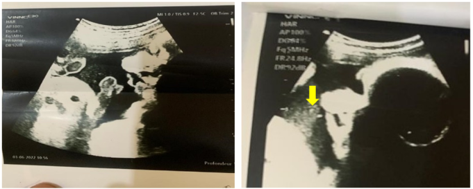
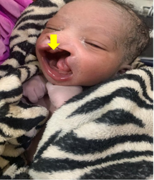
Introduction: A cleft lip or palate is a congenital malformation that causes a fissure of the lips or palate, and an antenatal diagnosis is essential for its treatment. Objective: The objective of this study was to present a case of a labial cleft in prenatal diagnosis by ultrasound at the Kalaban Coro reference health center. Materials and Methods: This was a case study of a labial cleft discovered during an obstetrical ultrasound. The examination was performed using a VINNO E30 ultrasound machine. Informed consent was obtained from the patient before her participation in the study. Participation in the study was voluntary. The patient's anonymity and confidentiality were guaranteed. Results: The study focused on a 28-year-old housewife. This was her third pregnancy and second delivery, with a history of stillbirth. No medical or surgical history was found. The patient was referred for an obstetrical ultrasound to evaluate the prognosis of delivery. The examination revealed a 34-week-old intrauterine male fetus with a large cleft lip and palate with umbilical cord interposition in the cleft, which was confirmed after birth. Conclusion: Ultrasound is the method of choice for prenatal screening of cleft lip and palate.
| Published in | Science Journal of Clinical Medicine (Volume 15, Issue 2) |
| DOI | 10.11648/j.sjcm.20261502.12 |
| Page(s) | 11-15 |
| Creative Commons |
This is an Open Access article, distributed under the terms of the Creative Commons Attribution 4.0 International License (http://creativecommons.org/licenses/by/4.0/), which permits unrestricted use, distribution and reproduction in any medium or format, provided the original work is properly cited. |
| Copyright |
Copyright © The Author(s), 2026. Published by Science Publishing Group |
Diagnosis, Ultrasound, Antepartum, Labial Cleft, Bamako, Mali
BPD | Biparietal |
FL | Femur Length |
FLP | Cleft Lip and Palate |
MRI | Magnetic Resonance Imaging |
CA | Abdominal Circumference |
HC | Head Circumference |
| [1] | McLeod NMH, Urioste MLA, Saeed NR. Birth prevalence of cleft lip and palate in Sucre, Bolivia. Cleft Palate Craniofac J. mars 2004; 41(2): 195‑8. |
| [2] | Szyszka-Sommerfeld L, Woźniak K, Matthews-Brzozowska T, Kawala B, Mikulewicz M, Machoy M. The electrical activity of the masticatory muscles in children with cleft lip and palate. Int J Paediatr Dent. mars 2018; 28(2): 257‑65. |
| [3] | Rekawek P, Coleman BG, Kamath A, Stone JL. Prenatal sonography of multicentric infantile myofibromatosis: Case report and review of the literature. J Clin Ultrasound. oct 2019; 47(8): 490‑3. |
| [4] | Davalbhakta A, Hall PN. The impact of antenatal diagnosis on the effectiveness and timing of counselling for cleft lip and palate. Br J Plast Surg. juin 2000; 53(4): 298‑301. |
| [5] | Heinrich A, Proff P, Michel T, Ruhland F, Kirbschus A, Gedrange T. Prenatal diagnostics of cleft deformities and its significance for parent and infant care. J Craniomaxillofac Surg. sept 2006; 34 Suppl 2: 14‑6. |
| [6] | Zheng W, Li B, Zou Y, Lou F. The prenatal diagnosis and classification of cleft palate: the role and value of magnetic resonance imaging. Eur Radiol. oct 2019; 29(10): 5600‑6. |
| [7] | Arangio P, Manganaro L, Pacifici A, Basile E, Cascone P. Importance of fetal MRI in evaluation of craniofacial deformities. J Craniofac Surg. mai 2013; 24(3): 773‑6. |
| [8] | Salari N, Darvishi N, Heydari M, Bokaee S, Darvishi F, Mohammadi M. Global prevalence of cleft palate, cleft lip and cleft palate and lip: A comprehensive systematic review and meta-analysis. J Stomatol Oral Maxillofac Surg. avr 2022; 123(2): 110‑20. |
| [9] | Tolarová MM, Cervenka J. Classification and birth prevalence of orofacial clefts. Am J Med Genet. 13 janv 1998; 75(2): 126‑37. |
| [10] | Lv M, Zhao B, Luo Q. Prenatal diagnosis and prognosis assessment of fetal intra-abdominal cystic lesions: a retrospective study in 264 cases. J Obstet Gynaecol. oct 2019; 39(7): 922‑7. |
| [11] | Al Omari F, Al-Omari IK. Cleft lip and palate in Jordan: birth prevalence rate. Cleft Palate Craniofac J. nov 2004; 41(6): 609‑12. |
| [12] | Moussa M, Abba Kaka HY, Roufai L, Eboungabeka Trigo ER, Bancole Pognon SA, Negrini JP. Cleft Lip and Palate in Niger: Epidemiological, Clinical, Therapeutic, and Long-Term Outcomes in 285 Cases . Health Sciences & Disease. 2020 ; 21 (10) : 18-22. |
| [13] | Dramé., B. M., Sanogo, A., Bah, A., Kokaina., C., TRAORE, T., Cissé., O., Camara, M., Beye, S. A., & TRAORE, H. (2018). The management of labial folds at the Nianankoro Fomba Hospital in Segou. Revue Malienne of Science and Technology, (20), 59–71. |
| [14] | Agrawal K. Cleft palate repair and variations. Indian J Plast Surg. oct 2009; 42(Suppl): S102‑9. |
| [15] | Guichoud Y, El Ezzi O, de Buys Roessingh A. Cleft Lip and Palate Antenatal Diagnosis: A Swiss University Center Performance Analysis. Diagnostics (Basel). 26 juill 2023; 13(15): 2479. |
| [16] | Brăila AD, Damian CM, Albu CC, Botoacă O, Dȋră LM, Albu ŞD, et al. Prenatal Diagnosis of Cleft Lip and Palate: A Retrospective Study. J Clin Med. 15 août 2024; 13(16): 4804. |
| [17] | Shao X., Liang L., Liu Y., Yan J., Ma P. Comparison of diagnostic values between 2D three-section ultrasound and 3D tomographic ultrasound imaging for fetal cleft palate at 11–13+6 weeks. J. Radiat. Res. Appl. Sci. 2024; 17: 100808. |
| [18] | Văduva CC, Constantinescu C, Ţenovici M, Văduva AR, Niculescu M, DiŢescu D, et al. Delayed interval delivery in twin pregnancy - case reports. Rom J Morphol Embryol. 2016; 57(3): 1089‑98. |
| [19] | Zhu Y, Miao H, Zeng Q, Li B, Wang D, Yu X, et al. Prevalence of cleft lip and/or cleft palate in Guangdong province, China, 2015–2018: a spatio-temporal descriptive analysis. BMJ Open. 2 août 2021; 11(8): e046430. |
| [20] | Yan X, Xing G, Wang X, Li J, Sun Q, Shang X. Diagnostic Value and Application of Prenatal MRI and Ultrasound in Fetal Cleft Lip and Palate. Contrast Media Mol Imaging. 18 mai 2022; 2022: 9410161. |
| [21] | Requeijo M. J. R., Penoni K. Z., Ruano R., Bunduki V., Lopes M. A. B., Francisco R. P. V., Zugaib M. Facial cleft diagnosed prenatally: Epidemiological, ultrasound and postnatal aspects. Ibero-American Journal of Humanities, Sciences, and Education. 2024; 10: 650–668. |
| [22] | Stoll C, Alembik Y, Dott B, Roth MP. Epidemiological and genetic study in 207 cases of oral clefts in Alsace, north-eastern France. J Med Genet. mai 1991; 28(5): 325‑9. |
| [23] | Milan M, Astolfi G, Volpato S, Garani GP, Clementi M, Tenconi R, et al. 766 cases of oral cleft in Italy. Data from Emilia Romagna (IMER) and northeast Italy (NEI) registers. Eur J Epidemiol. juin 1994; 10(3): 317‑24. |
| [24] | Martelli DRB, da Cruz KW, de Barros LM, Silveira MF, Oliveira Swerts MS, Júnior HM. Maternal and paternal age, birth order and interpregnancy interval evaluation for cleft lip-palate. Braz J Otorhinolaryngol. 17 oct 2015; 76(1): 107‑12. |
APA Style
Abdoulaye, C. M., Traore, M. S., Yanogue, A., Sidibe, B., Traore, B., et al. (2026). Prenatal Ultrasound Diagnosis of Cleft Lip and Palate: A Case Report. Science Journal of Clinical Medicine, 15(2), 11-15. https://doi.org/10.11648/j.sjcm.20261502.12
ACS Style
Abdoulaye, C. M.; Traore, M. S.; Yanogue, A.; Sidibe, B.; Traore, B., et al. Prenatal Ultrasound Diagnosis of Cleft Lip and Palate: A Case Report. Sci. J. Clin. Med. 2026, 15(2), 11-15. doi: 10.11648/j.sjcm.20261502.12
@article{10.11648/j.sjcm.20261502.12,
  author = {Camara Mody Abdoulaye and Mamadou Sylla Traore and Aldjouma Yanogue and Bamody Sidibe and Bagnoni Traore and Issa Traore and Guindo Ilias and Tata Toure and Siaka Sidibe},
  title = {Prenatal Ultrasound Diagnosis of Cleft Lip and Palate:
A Case Report},
  journal = {Science Journal of Clinical Medicine},
  volume = {15},
  number = {2},
  pages = {11-15},
  doi = {10.11648/j.sjcm.20261502.12},
  url = {https://doi.org/10.11648/j.sjcm.20261502.12},
  eprint = {https://article.sciencepublishinggroup.com/pdf/10.11648.j.sjcm.20261502.12},
  abstract = {Introduction: A cleft lip or palate is a congenital malformation that causes a fissure of the lips or palate, and an antenatal diagnosis is essential for its treatment. Objective: The objective of this study was to present a case of a labial cleft in prenatal diagnosis by ultrasound at the Kalaban Coro reference health center. Materials and Methods: This was a case study of a labial cleft discovered during an obstetrical ultrasound. The examination was performed using a VINNO E30 ultrasound machine. Informed consent was obtained from the patient before her participation in the study. Participation in the study was voluntary. The patient's anonymity and confidentiality were guaranteed. Results: The study focused on a 28-year-old housewife. This was her third pregnancy and second delivery, with a history of stillbirth. No medical or surgical history was found. The patient was referred for an obstetrical ultrasound to evaluate the prognosis of delivery. The examination revealed a 34-week-old intrauterine male fetus with a large cleft lip and palate with umbilical cord interposition in the cleft, which was confirmed after birth. Conclusion: Ultrasound is the method of choice for prenatal screening of cleft lip and palate.},
 year = {2026}
}
TY  - JOUR
T1  - Prenatal Ultrasound Diagnosis of Cleft Lip and Palate:
A Case Report
AU  - Camara Mody Abdoulaye
AU  - Mamadou Sylla Traore
AU  - Aldjouma Yanogue
AU  - Bamody Sidibe
AU  - Bagnoni Traore
AU  - Issa Traore
AU  - Guindo Ilias
AU  - Tata Toure
AU  - Siaka Sidibe
Y1  - 2026/04/30
PY  - 2026
N1  - https://doi.org/10.11648/j.sjcm.20261502.12
DO  - 10.11648/j.sjcm.20261502.12
T2  - Science Journal of Clinical Medicine
JF  - Science Journal of Clinical Medicine
JO  - Science Journal of Clinical Medicine
SP  - 11
EP  - 15
PB  - Science Publishing Group
SN  - 2327-2732
UR  - https://doi.org/10.11648/j.sjcm.20261502.12
AB  - Introduction: A cleft lip or palate is a congenital malformation that causes a fissure of the lips or palate, and an antenatal diagnosis is essential for its treatment. Objective: The objective of this study was to present a case of a labial cleft in prenatal diagnosis by ultrasound at the Kalaban Coro reference health center. Materials and Methods: This was a case study of a labial cleft discovered during an obstetrical ultrasound. The examination was performed using a VINNO E30 ultrasound machine. Informed consent was obtained from the patient before her participation in the study. Participation in the study was voluntary. The patient's anonymity and confidentiality were guaranteed. Results: The study focused on a 28-year-old housewife. This was her third pregnancy and second delivery, with a history of stillbirth. No medical or surgical history was found. The patient was referred for an obstetrical ultrasound to evaluate the prognosis of delivery. The examination revealed a 34-week-old intrauterine male fetus with a large cleft lip and palate with umbilical cord interposition in the cleft, which was confirmed after birth. Conclusion: Ultrasound is the method of choice for prenatal screening of cleft lip and palate.
VL  - 15
IS  - 2
ER  -
Department of Radiology and Medical Imaging, Professor Bocar Sidy Sall University Hospital, Kati, Mali
Information